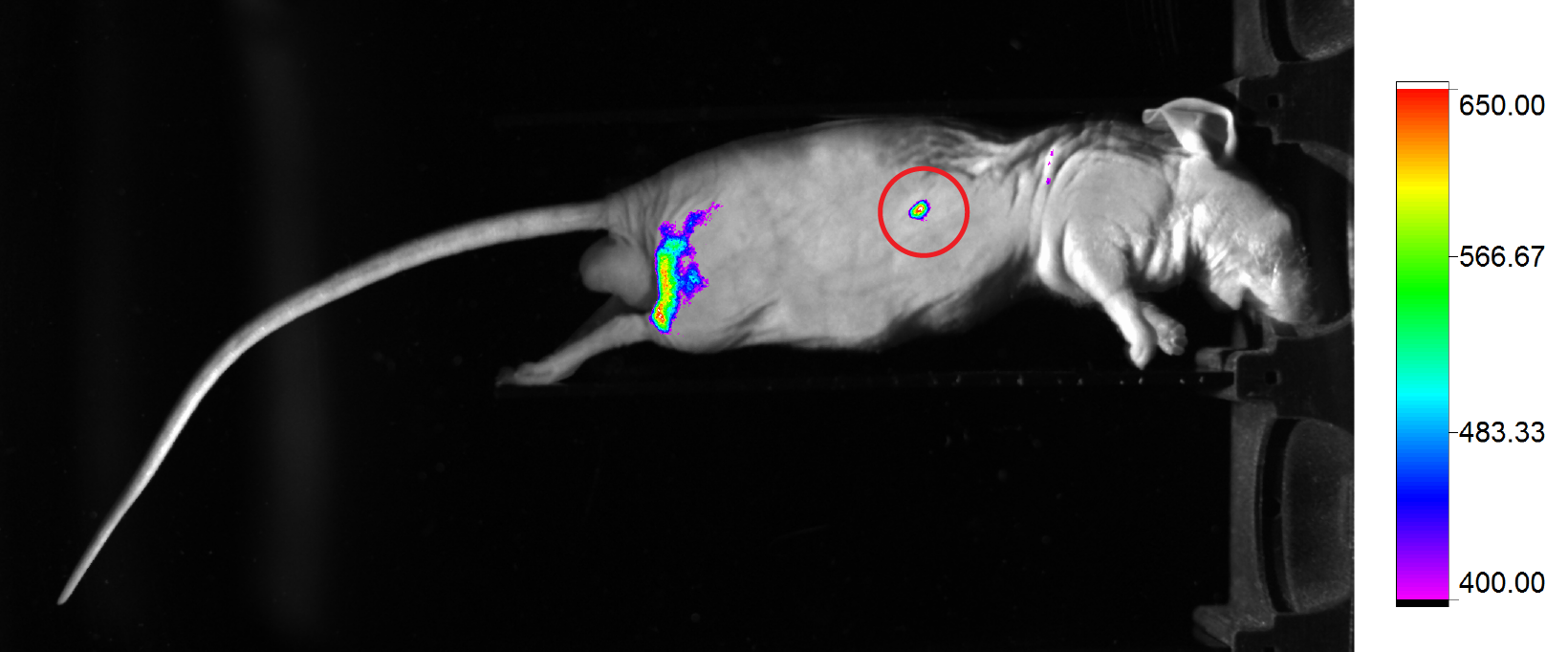
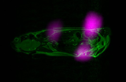
In-vivo imaging of small laboratory animals includes both anatomical/morphological, and functional/molecularimaging techniques for imaging live animals for research purposes. The most common anatomical imaging methods such as magnetic resonance imaging (MRI), computed tomography (CT), and high frequency ultrasound provide high resolution images of internal structures of the body (bone, soft tissues). They provide information about the size, morphology and structural changes of individual organs, but they tell us little about the functional or metabolic activity of tissue. The most commonly used methods for molecular imaging are positron emission tomography (PET), single photon emission computed tomography (SPECT), and optical imaging (fluorescence and bioluminescence). Recently, a promising molecular imaging method has been developed based on direct detection of the paramagnetic contrast: magnetic particle imaging (MPI). Molecular imaging methods have high sensitivity and specificity but in comparison with anatomical imaging techniques, they offer considerably lower spatial resolution and they do not reveal precise anatomical details. Recent advances in instrumentation and development of imaging contrast agents for multimodal imaging both anatomical and molecular imaging approaches to be linked in an advantageous manner.
 |
 |
 |
Molecular imaging is gaining importance for the direct visualization of specific molecular processes in vivo, especially those which are specific for different diseases. Several imaging techniques that can be considered to be molecular were developed several decades ago (e.g., imaging using labeled monoclonal antibodies and radioisotope imaging of receptors, etc..). Only recently have the necessary tools that enable us to significantly improve imaging results become available. These tools include molecular cloning, nanotechnology, robotics, crystallography, fast mass spectrometry, and sophisticated computer analysis. New and improved methods will allow us to explore more basic biological questions in vivo.
Several key criteria should be fulfilled to view a specific molecule in vivo:
- the availability of suitable probes with high affinity and reasonable pharmacodynamics
- the ability of these probes to overcome biological barriers (intestinal or vascular wall, cell membrane)
- accumulation of the probe in the target tissue (chemical or biological signal amplification)
- the availability of imaging methods with high sensitivity and resolution and sufficient speed
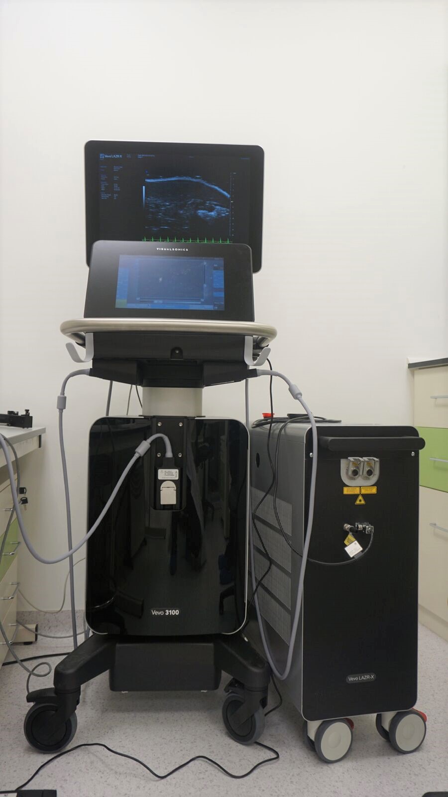
VEVO LAZR-X ultrasound, photoacoustics
|
BRUKER ICON 1T |
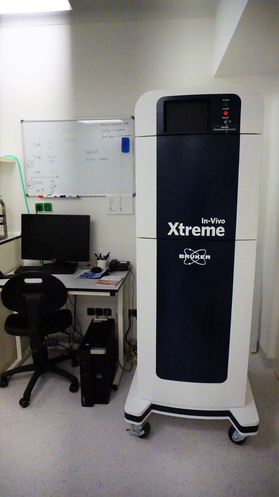
BRUKER Xtreme X- ray / Optical Imaging |
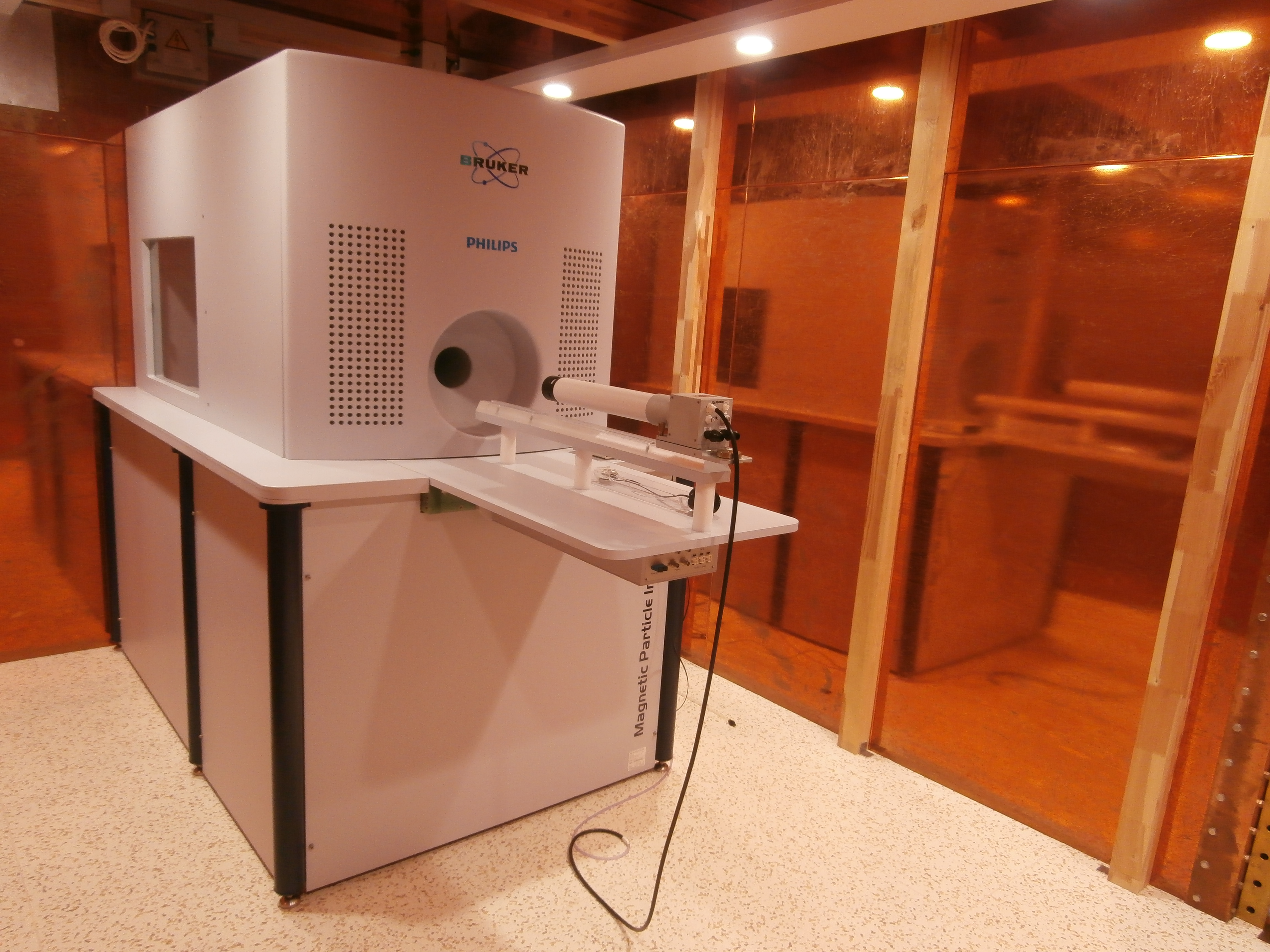
BRUKER MPI - Magnetic Particle Imaging
|
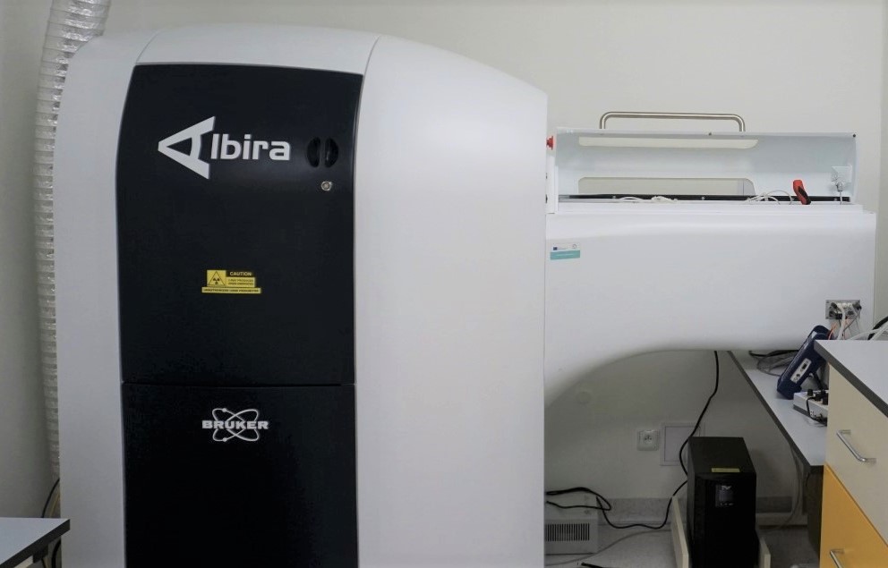
BRUKER Albira CT/ PET/ SPECT
|
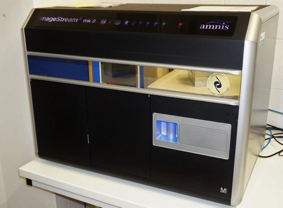
AMNIS Imaging Flow Cytometer
|
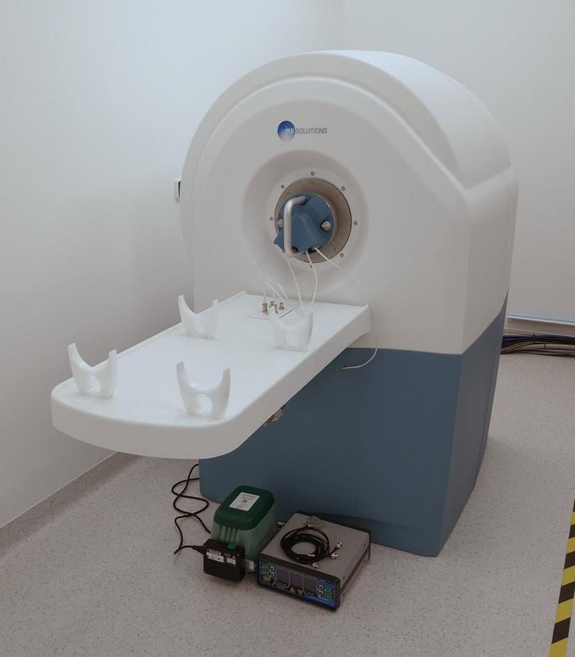
7 T MRI (MR Solutions) |
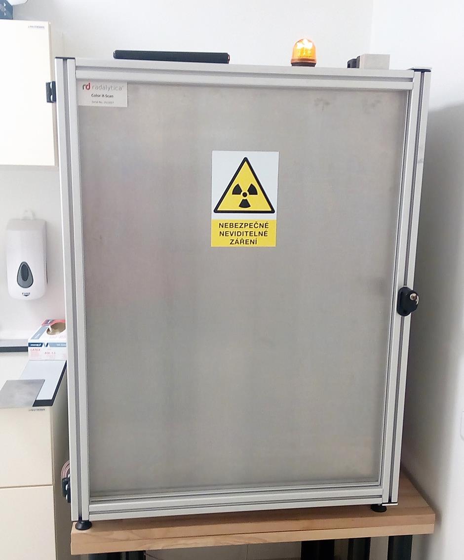
2D spectral X-ray Color- X- Scan
|