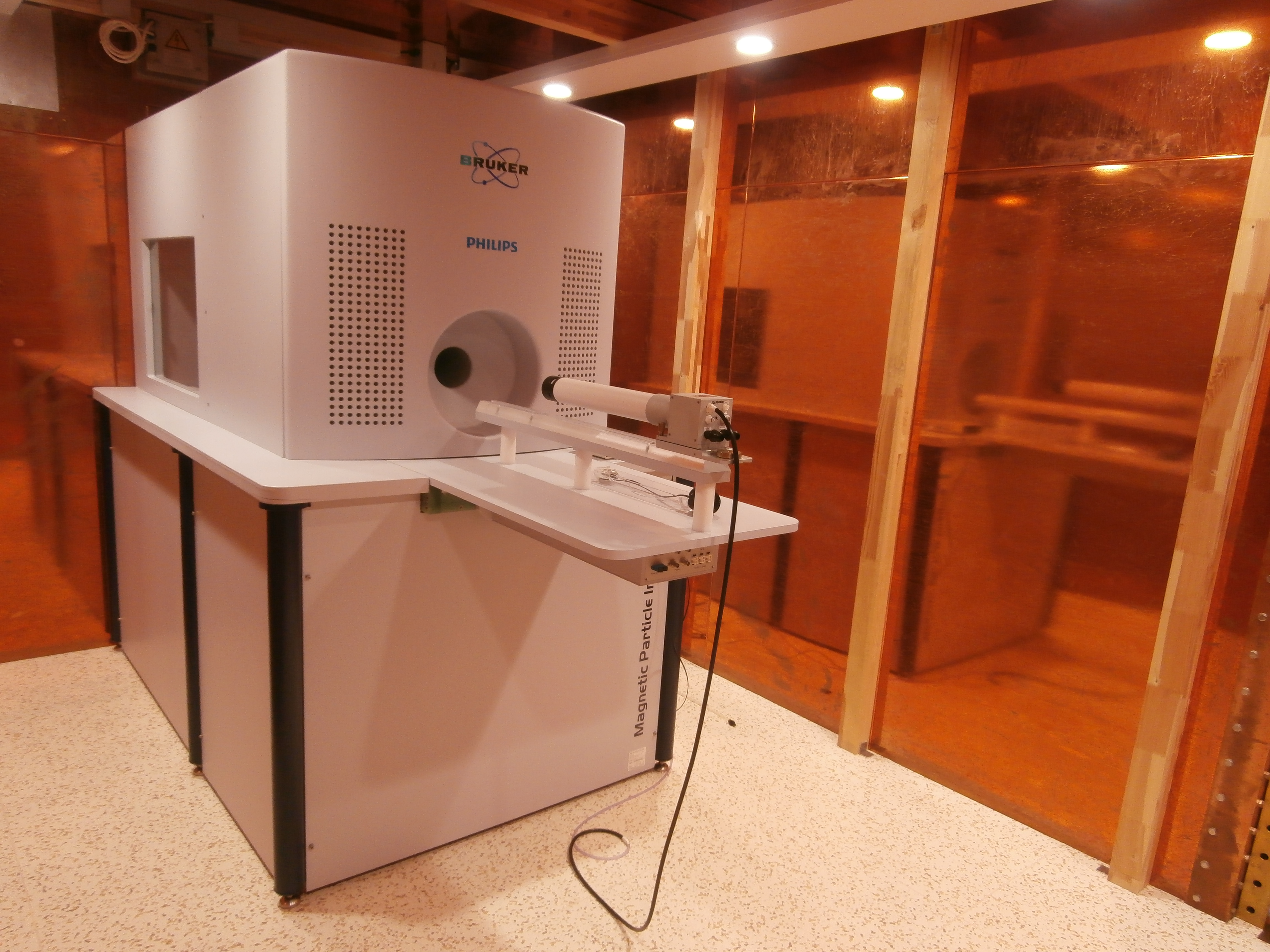
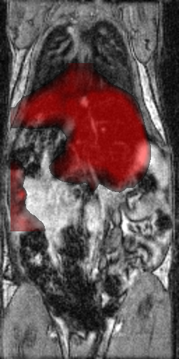
MPI (Bruker Biospin, Ettlingen, Germany) enables direct detection of paramagnetic nanoparticles in an oscillating magnetic field. Magnetic particle detection is a quantitative (no attenuation of the signal) tomographic 3D functional imaging technique with a high scan rate (up to 46 images/s). In combination with MRI or CT, this enables high resolution sensitive monitoring of superparamagnetic nanoparticles (SPIO, super paramagnetic iron oxide nanoparticles) that can visualize the bloodstream and to track cells or macromolecules, monitor their movements and distribution in an organism. MPI is 1.000 times faster than PET and 100 times more sensitive than MRI
Magnetic field gradient: 2.5 T / m Applications: · Nontoxic angiography MPI Magnetic Particle Imager Brochure
Basic Technical Parameters:
Acquisition speed: 20 ms
Spatial resolution <0.6 mm
Sensitivity: 10-11 mol / l
Rapid quantitative imaging of magnetic contrast
· Cell tracking
· Research on drug carriers
· Nano-robotics
· Cardiovascular research
· Regenerative medicine